Overview
Do you experience pain at the back of your heel? Is the back of your heel red and swollen? And you were told that it might Achilles Tendonitis? But so far treatment for Achilles Tendonitis does not seem to be working? You might be suffering instead from Retrocalcaneal Bursitis. Causes There are several factors which can lead to a person developing retrocalcaneal bursitis. In athletes, especially runners, overtraining, sudden excessive increase in running mileage may lead to retrocalcaneal bursitis. Tight or ill-fitting shoes can be another causative factor as they can produce excessive pressure at the back of the heel due to restrictive heel counter. A person with an excessively prominent posterosuperior aspect of the heel bone (Haglund deformity) may also have a higher predisposition to retrocalcaneal bursitis. In such individuals, pain would be reproduced when the ankle goes into dorsiflexion. Symptoms A person with bursitis can have one or more of the symptoms below. Pain, the pain increases with movement or pressure. Tenderness is felt even without movement. Swelling. Loss of movement. If the bursitis is caused by an infection it is called Septic Bursitis. The patient with septic bursitis may have the following additional symptoms. Fever. The affected area is red. The affected area feels hot when touched. Diagnosis Obtaining a detailed history from the patient is important in diagnosing calcaneal bursitis. The following complaints (which the physician should ask about during the subjective examination) are commonly reported by patients. Other inquiries that the physician should make include the following. The clinician should ask about the patient's customary footwear (whether, for example, it includes high-heeled shoes or tight-fitting athletic shoes). The patient should be asked specifically about any recent change in footwear, such as whether he/she is wearing new athletic shoes or whether the patient has made a transition from flat shoes to high heels or vice versa. Individuals who have been accustomed to wearing high-heeled shoes on a long-term basis may find that switching to flat shoes causes increased stretch and irritation of the Achilles tendon and the associated bursae. The specifics of a patient's activity level should be ascertained, including how far the patient runs and, in particular, whether the individual is running with greater intensity than before or has increased the distance being run. The history of any known or suspected underlying rheumatologic conditions, such as gout, rheumatoid arthritis, or seronegative spondyloarthropathies, should be obtained. Non Surgical Treatment Podiatric Care may include using anti-inflammatory oral medications or an injection of medication and local anesthetic to reduce the swelling in the bursa. An injection may be used for both diagnosis and for treatment. When you go to your doctor, x-rays are usually required to evaluate the structure of your foot and ankle to ensure no other problems exist in this area. They may advise you on different shoewear or prescribe a custom made orthotic to try and control the foot structure especially if you have excessive pronation. Sometimes patients are sent to Physical Therapy for treatment as well. To aid in relief of pressure points, some simple padding techniques can be utilized. Most all patients respond to these conservative measures once the area of irritation is removed. Surgical Treatment Surgery is rarely need to treat most of these conditions. A patient with a soft tissue rheumatic syndrome may need surgery, however, if problems persist and other treatment methods do not help symptoms. Prevention People can lower the risk of bursitis by gradually strengthening and stretching the muscles around the joints and taking regular breaks from repetitive motion that might irritate bursae. Prolonged time resting on the elbows or kneeling should be avoided, if it cannot be avoided, wearing cushioned elbow and knee pads can help protect the bursae. Comfortable, supportive, low-heeled shoes can help prevent bursitis in the foot.  Overview OverviewHammer toes is often a harmless and painless condition. Although the toe may be curled permanently, hammertoe should not cause any long-term problems other than a more difficult time finding shoes that fit. If hammertoe is treated and preventive measures are followed, the condition should not return. Wearing tight or constricting shoes can cause hammertoe to return. Causes Your toe contains two joints that allow it to bend at the middle and bottom. A hammertoe occurs when the middle joint becomes dislocated. Common causes of this joint dislocation include a toe injury, arthritis, a high foot arch, wearing shoes that don?t fit properly, tightened ligaments or tendons in the foot, pressure from a bunion (when your big toe points inward toward your second toe) Spinal cord or peripheral nerve damage may cause all of your toes to curl downward.  Symptoms SymptomsA hammertoe may be hammertoe present but not always painful unless irritated by shoes. One may have enlarged toe joints with some thickened skin and no redness or swelling. However, if shoes create pressure on the joint, the pain will usually range from pinching and squeezing to sharp and burning. In long standing conditions, the dislocated joints can cause the pain of arthritis. Diagnosis Hammertoes are progressive, they don?t go away by themselves and usually they will get worse over time. However, not all cases are alike, some hammertoes progress more rapidly than others. Once your foot and ankle surgeon has evaluated your hammertoes, a treatment plan can be developed that is suited to your needs. Non Surgical Treatment Changing the type of footwear worn is a very important step in the treatment of hammer toes. When choosing a shoe, make sure the toe box (toe area) is high and broad, and can accommodate the hammer toes. A shoe with a high, broad toe box will provide enough room in the forefoot area so that there is less friction against the toes. Other conservative treatments include using forefoot products designed to relieve hammer toes, such as hammer toe crests and hammer toe splints. These devices will help hold down the hammer toe and provide relief to the forefoot. Gel toe shields and gel toe caps are also recommended to eliminate friction between the shoe and the toe, while providing comfort and lubrication. Surgical Treatment Surgery may not help how your foot looks. And your toe problems may also come back after surgery. This is more likely if you keep wearing the kinds of shoes that cause toe problems. Your expectations will play a large role in how you feel about the results of surgery. If you are only having surgery to improve the way your foot looks, you may not be happy with how it turns out.  Overview OverviewHammer toes is often a harmless and painless condition. Although the toe may be curled permanently, hammertoe should not cause any long-term problems other than a more difficult time finding shoes that fit. If hammertoe is treated and preventive measures are followed, the condition should not return. Wearing tight or constricting shoes can cause hammertoe to return. Causes The main cause of hammer toe is poorly fitted and/or poorly designed footwear. Any footwear that is too tight in the toe box, especially high-heeled shoes, can push the toes forward, crowding one or more of them into a space that is not large enough to allow the toes to lie flat and spread as they should. Other causes include the following. Changes in foot anatomy. Sometimes the metatarsal bones in the ball of the foot can ?drop,? creating a situation in which the toes do not make contact with the surface of the shoe. The toes may then contract at one or both of the joints to re-establish contact with the surface. Traumatic injuries in which toes are jammed or broken. Diabetic neuropathy. This can cause abnormal foot biomechanics due to nerve and/or muscle damage. Damage to nerves and muscles from other conditions, such as arthritis or stroke. Heredity. 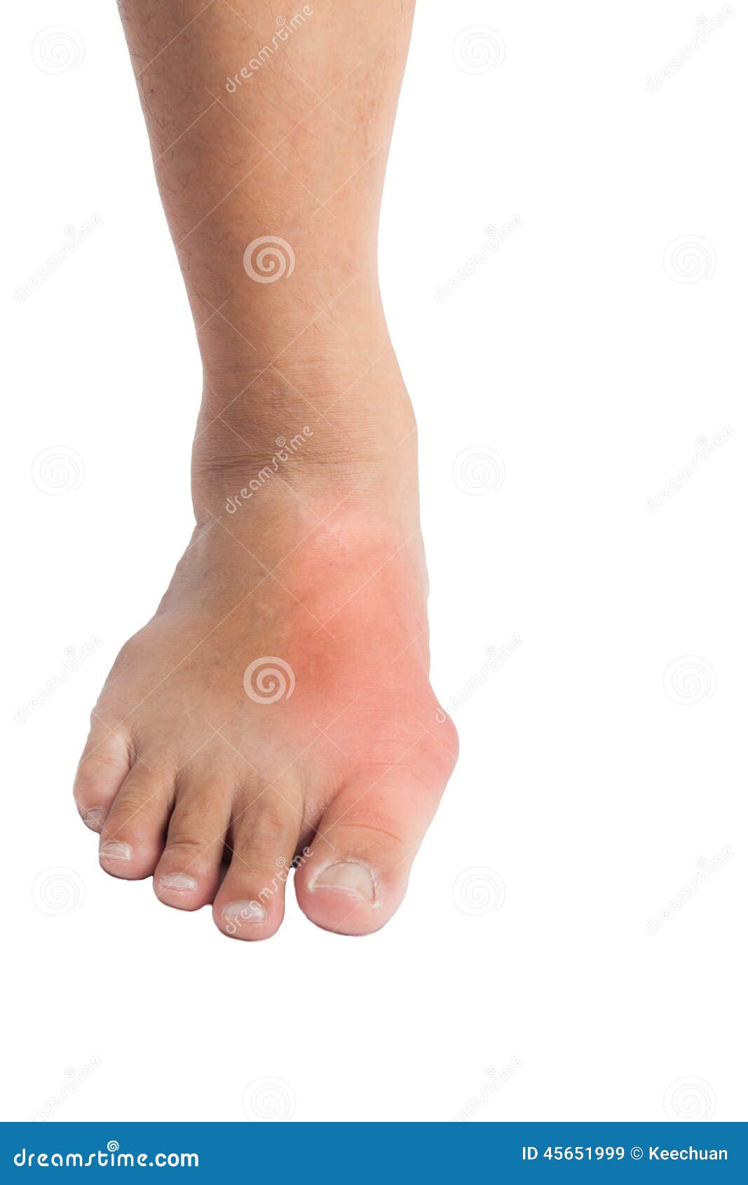 Symptoms SymptomsThe most common symptoms of hammertoes include. The toe is bent upward at the middle toe joint, so that the top of this joint rubs against the top of the shoe. The remainder of the toe is bent downward. Pain upon pressure at the top of the bent toe from footwear. The formation of corns on the top of the joint. Redness and swelling at the joint contracture. Restricted or painful motion of the toe joint. Pain in the ball of the foot at the base of the affected toe. This occurs because the contracted digit puts pressure on the metatarsal head creating callouse and pressure on the ball of the foot. Diagnosis Most health care professionals can diagnose hammertoe simply by examining your toes and feet. X-rays of the feet are not needed to diagnose hammertoe, but they may be useful to look for signs of some types of arthritis (such as rheumatoid arthritis) or other disorders that can cause hammertoe. Non Surgical Treatment A number of approaches can be undertaken to the manage a hammer toe. It is important that any footwear advice is followed. The correct amount of space in the toe box will allow room for the toes to function without excessive pressure. If a corn is present, this will need to be treated. If the toe is still flexible, it may be possible to use splints or tape to try and correct the toe. Without correct fitting footwear, this is often unsuccessful. Padding is often used to get pressure off the toe to help the symptoms. If conservative treatment is unsuccessful at helping the symptoms, surgery is often a good option. Surgical Treatment If these treatments are not sufficient at correcting the hammer toe, an operation to straighten the toe may be necessary. This is often performed in conjunction with surgery for a bunion deformity. The surgical treatment of a hammer hammertoe toe can consist of either cutting the tendons to relieve the pressure that causes the deformity, or fusing the toe so that it points straight permanently.  Prevention PreventionTo help prevent hammer toes from developing, wear shoes or boots that provide sufficient width in the toe box to ensure minimal compression. Use inserts that help the toes flatten out and spread and give sufficient support to the metatarsal arch in the forefoot. If hammer toes have already formed, padded socks help protect the tops and the tips of the hammer toes and may reduce pain from rubbing and chafing.
Overview
 Hallux valgus is a condition that affects the joint at the base of the big toe. The condition is commonly called a bunion. The bunion actually refers to the bump that grows on the side of the first metatarsophalangeal (MTP) joint. In reality, the condition is much more complex than a simple bump on the side of the toe. Interestingly, this condition almost never occurs in cultures that do not wear shoes. Pointed shoes, such as high heels and cowboy boots, can contribute to the development of hallux valgus. Wide shoes, with plenty of room for the toes, lessen the chances of developing the deformity and help reduce the irritation on the bunion if you already have one. Hallux valgus is a condition that affects the joint at the base of the big toe. The condition is commonly called a bunion. The bunion actually refers to the bump that grows on the side of the first metatarsophalangeal (MTP) joint. In reality, the condition is much more complex than a simple bump on the side of the toe. Interestingly, this condition almost never occurs in cultures that do not wear shoes. Pointed shoes, such as high heels and cowboy boots, can contribute to the development of hallux valgus. Wide shoes, with plenty of room for the toes, lessen the chances of developing the deformity and help reduce the irritation on the bunion if you already have one.Causes Bunions occur with greater regularity in women than men, and they may sometimes run in families. You may also have an increased likelihood of bunions if you are born with certain bone abnormalities in your feet. Factors that may increase your chances of developing a bunion include long-term use of narrow-toed and/or high-heeled footwear. Arthritis. Toe trauma. Laxity of your connective tissues (ligament laxity). Limb length inequalities. Genetics. Certain foot problems (e.g. flatfoot, over-pronation, etc.). Symptoms Movement of the big toe towards the smaller toes. Bulging bump on the outside of the base of the big toe. Restricted movement of the big toe. Swelling, inflammation, redness or soreness around your big toe joint. Persistent or sporadic dull, sharp or aching pain in or around the big toe. Corns, blisters and calluses which can develop when the first and second toes overlap. Over time, more severe symptoms can occur such as arthritis of the big toe, stress fractures and problems walking. Diagnosis Most patients are diagnosed to have bunions from clinical history and examination. However, in some cases, X-rays will be performed to determine the extent of damage to the joint. Furthermore, it will enable the treating doctor to decide on the best course of management of the patient. Non Surgical Treatment Several things can be done to help relive the pain of bunions. These won't make the bunion go away, but they can make the foot more comfortable. Wearing different shoes. Shoes with a wide toe box rather than a pointed one will help. Shoes with lower heels will also help. (High heels throw more of the body's weight on the front part of the foot where the toe joints are.) Padding. Pads placed over the bunion may help reduce the pain. These are available from a drug store or may be available from a foot and ankle surgeon. Avoiding activities that make the pain worse. This includes standing for a long time or other activities that make the bunion sore. Non-steroidal anti-inflammatory drugs. These include aspirin or ibuprofen. They relieve pain and swelling. Applying an ice pack to reduce swelling and pain. Corticosteroid injections. These are not often used in bunion treatment. Injecting corticosteroids sometimes helps if the bursa is inflamed. (Bursa is a fluid-filled sac within a joint to cushion the bones). Orthotic devices. These are devices placed inside a shoe that shift the positioning of the foot. Orthotics help compensate for structural issues that cause foot problems.  Surgical Treatment Conservative bunion treatment methods help to reduce the symptoms but they cannot undo changes in the bone to fully correct the condition. If the pain becomes severe and it starts to affect your daily life, then foot bunion surgery may be the best option to correct a hallux abducto valgus. There are a number of different surgical options, depending on the severity of the bony deformity. The two most common types of surgery are Osteotomy. This is the most common type pf surgery and involves removing part of the bony lump and realigning the toes. It is also known as a bunionectomy or exostectomy. The ligaments around the big toe may also need realigning and this will be done at the same time if necessary. Fusion.This is where the joint at the base of the big toe (metatarsophalangeal joint) is fused together, known as an arthrodesis. This is only indicated in severe cases or if other treatments have failed as it severely limits the movement of the big toe. Prevention The best way to reduce your chances of developing bunions is to wear shoes that fit properly. Shoes that are too tight or have high heels can force your toes together. Bunions are rare in populations that don?t wear shoes. Make sure your shoes are the correct size and that there's enough room to move your toes freely. It's best to avoid wearing shoes with high heels or pointed toes. Overview
Too much of a good thing can be bad, and when it comes to pronation of the foot this is often the case. When the foot rolls too much during contact with the ground, the body weight is not cushioned effectively. The degree to which pronation occurs may be difficult to spot without someone observing how you run. It is easier to tell if a runner is pronating excessively when they are observed from behind rather than from above. When overpronation is only mild to moderate it may not be easy to spot without having a professional gait analysis performed.  Causes Over-pronation is very prominent in people who have flexible, flat feet. The framework of the foot begins to collapse, causing the foot to flatten and adding additional stress to other parts of the foot. Therefore over-pronation can often lead to a case of Plantar Fasciitis, Heel Spurs, Metatarsalgia, Post-tib Tendonitis and/or Bunions. There are many causes of flat feet. Obesity, pregnancy or repetitive pounding on a hard surface can weaken the arch leading to over-pronation. People with flat feet often do not experience discomfort immediately, and some never suffer from any discomfort at all. However, when symptoms develop and become painful, walking becomes awkward and causes increased strain on the feet and calves. Symptoms Overpronation can lead to injuries and pain in the foot, ankle, knee, or hip. Overpronation puts extra stress on all the bones in the feet. The repeated stress on the knees, shins, thighs, and pelvis puts additional stress on the muscles, tendons, and ligaments of the lower leg. This can put the knee, hip, and back out of alignment, and it can become very painful. Diagnosis Look at the wear on your shoes and especially running trainers; if you overpronate it's likely the inside of your shoe will be worn down (or seem crushed if they're soft shoes) from the extra strain.  Non Surgical Treatment Not all over pronation is treated. Although, when it appears to be a causitive factor that is contributing to pain,or development of structural deformities, there are various degrees of treatment.In some cases specific shoes may be all that is required. In other cases, paddings or strapping, are prescribed and where necessary orthotic therapy. A podiatric assesment would be advised to asses this. Surgical Treatment HyProCure implant. A stent is placed into a naturally occurring space between the ankle bone and the heel bone/midfoot bone. The stent realigns the surfaces of the bones, allowing normal joint function. Generally tolerated in both pediatric and adult patients, with or without adjunct soft tissue procedures. Reported removal rates, published in scientific journals vary from 1%-6%. Overview
Sever's disease is a term used to describe inflammation of the calcaneal apophysis which occurs in children and adolescents. Sever first described the condition in 1912. Further studies have suggested that the condition is due to repeated 'microtrauma' at the site of the attachment of the Achilles tendon to the apophysis of the heel, often as result of sporting activities. The disorder can be classified among the general osteochondrosis syndromes such as Osgood-Schlatter disease. Causes Sever?s disease is most likely to occur during the growth spurt that occurs in adolescence. For girls, growth spurts usually occurs between 8 and 13 years of age. For boys, it?s typically between 10 and 15 years of age. The back of the heel hardens and becomes stronger when it finishes growing, which is why Sever?s rarely occurs in older adolescents and teenagers. Symptoms The symptoms of Sever?s disease occur in the heel and the foot, and may worsen with activity. Pain and stiffness can occur in one or both heels. Symptoms can include. Swelling in the heel. Redness in the heel. Antalgic gait (such as limping). Foot pain or stiffness first thing in the morning or while walking. Pain that is worsened by squeezing the heel. Diagnosis All medical diagnosis should be made by taking a full history, examining the patient then performing investigations. The problem usually occurs in boys who are going through or have just gone through a growth spurt; one or both heels may be affected. Initially the pain may be intermittent occurring only during or after exercise. As the problem gets worse, pain may be present most of the time. There may be swelling over the back of the heel and this area is painful if touched or knocked. On examination the patient often has flat feet, very tight legs muscles especially the gastrocnemius. Non Surgical Treatment Treatment for Sever?s disease is mainly supportive, to stop inflammation and reduce pain. The condition will resolve on its own when the growth in the growth plate is complete, but until then, measures can be taken to resolve pain and discomfort. Applying ice to the painful or swollen areas on the foot may provide some short-term relief from pain and prevent further inflammation. Ice can be applied for about 20 minutes two or three times a day. Footwear that is too big, too small, or does not provide proper support can exacerbate the symptoms of Sever?s disease. Supportive footwear is important to prevent discomfort, especially in children who participate in sports and activities that take place on a hard surface (such as pavement or a basketball court). Shoes should also have adequate padding and not rub against the heel. In some cases, shoes that do not have heels (such as sandals) may be more comfortable to wear while the heel is healing, but care should be taken that the shoe provides proper support to the rest of the foot. Children with Sever?s disease should avoid going barefoot.Children with flat feet, high arches, or over-pronation may need treatment to resolve these underlying conditions. In many cases, an orthotic worn inside the shoe can help put the foot into a better alignment and provide relief to the foot or the arch. Children who are overweight or obese may be counseled to lose weight. Being overweight can contribute to the development of several conditions, including Sever?s disease. Resting the foot and discontinuing sports and other activities until the pain and stiffness is resolved may be recommended. In extreme cases, a walking boot or a cast might be used to completely immobilize the foot. A physical therapist may recommend stretching exercises for the muscles in the calf and the Achilles tendon. A stretching routine is usually done several times a day. Stretching these muscles can help improve strength and decrease the stress on the heel plate. Some physicians may recommend over-the-counter pain relievers such as ibuprofen or acetaminophen. Care must be taken when administering these medications to children, especially with acetaminophen, as overdoses are possible when using more than one medication containing acetaminophen. Aspirin should never be given to children. The utility of pain relievers in children must be weighed against their possible side effects. For small variations-less than an inch or so-shoe lifts can help equalize the length of the legs. In cases with more variation between legs, surgical solutions may be considered. Research indicates that targeted manual therapy techniques performed by a licensed physical therapist can help to reduce pain from Sever?s Disease and to improve muscle function. When the larger calf muscles and the smaller ankle and foot muscles become tight, this tightness can affect the mechanics of the ankle joint. Manual therapy includes both joint and muscle release techniques to restore optimal function to the calf, ankle, and foot muscles, and results can generally be achieved within a few months. Prevention Sever?s disease may be prevented by maintaining good flexibility while your child is growing. The stretching exercises mentioned here can lower your child?s risk for injuries during the growth spurt. Good-quality shoes with firm support and a shock-absorbent sole will help. Your child should avoid excessive running on hard surfaces. If your child has already recovered from Sever?s disease, stretching and putting ice on the heel after activity will help keep your child from getting this condition again. Overview
 Heel Pain is a very challenging problem as it can be local and/or referred. It has been more prevalent recently due to the hard grounds on which people have to run. Commonly people will present with heel pain, thrusting an x-ray at you, and being adamant that the problem is a heel spur. This is defined as a small bone that grows from the heel, directing forwards towards the toes. This may be as small as 1 mm to anything as large as 8 - 10 mm. Most of the time, this is an incidental finding, as there many heels that are pain free that have heel spurs evident on x-rays. The spur is thought to be a result of traction of the plantar fascia on the heel. In some cases, the spur may contribute to the symptoms, but is not the main cause. This should be explained very carefully to the patient, as the focus on the spur may limit the recovery, as the patient may believe that the only way to eliminate the pain is to remove the spur. Causes In our pursuit of healthy bodies, pain can be an enemy. In some instances, however, it is of biological benefit. Pain that occurs right after an injury or early in an illness may play a protective role, often warning us about the damage we've suffered. When we sprain an ankle, for example, the pain warns us that the ligament and soft tissues may be frayed and bruised, and that further activity may cause additional injury. Pain, such as may occur in our heels, also alerts us to seek medical attention. This alert is of utmost importance because of the many afflictions that contribute to heel pain. Symptoms See your doctor immediately if you have Severe pain and swelling near your heel. Inability to bend your foot downward, rise on your toes or walk normally. Heel pain with fever, numbness or tingling in your heel. Severe heel pain immediately after an injury. Schedule an office visit if you have. Heel pain that continues when you're not walking or standing. Heel pain that lasts more than a few weeks, even after you've tried rest, ice and other home treatments. Diagnosis The diagnosis of plantar fasciitis is generally made during the history and physical examination. There are several conditions that can cause heel pain, and plantar fasciitis must be distinguished from these conditions. Pain can be referred to the heel and foot from other areas of the body such as the low back, hip, knee, and/or ankle. Special tests to challenge these areas are performed to help confirm the problem is truly coming from the plantar fascia. An X-ray may be ordered to rule out a stress fracture of the heel bone and to see if a bone spur is present that is large enough to cause problems. Other helpful imaging studies include bone scans, MRI, and ultrasound. Ultrasonographic exam may be favored as it is quick, less expensive, and does not expose you to radiation. Laboratory investigation may be necessary in some cases to rule out a systemic illness causing the heel pain, such as rheumatoid arthritis, Reiter's syndrome, or ankylosing spondylitis. These are diseases that affect the entire body but may show up at first as pain in the heel. Non Surgical Treatment When consulting a doctor about heel pain, a patient can expect to be questioned about their level of pain, how long they?ve been experiencing it, and which activities aggravate or alleviate the condition. The doctor may order x-rays, a physical therapy regimen, or refer the afflicted individual to an orthopedic specialist for further examination. The doctor may attempt to recreate conditions that cause the heel pain to flare up in order to study reaction and cause in the patient, but this will be temporary and the doctor will stop this test at the request of the patient. A patient with heel pain may also be fitted with special inserts for their shoes to help correct arch and heel problems that cause pain. Heel problems can range from the mildly irritating to the nearly devastating, but proper prevention in care will help keep each step pain free. Advanced orthopedics, pain management, and technologically-honed surgical techniques ensure that no patient needs to suffer with the discomfort of heel pain and the restrictions it imposes on an active lifestyle. Surgical Treatment If treatment hasn't worked and you still have painful symptoms after a year, your GP may refer you to either an orthopaedic surgeon, a surgeon who specialises in surgery that involves bones, muscles and joints or a podiatric surgeon, a podiatrist who specialises in foot surgery. Surgery is sometimes recommended for professional athletes and other sportspeople whose heel pain is adversely affecting their career. Plantar release surgery is the most widely used type of surgery for heel pain. The surgeon will cut the fascia to release it from your heel bone and reduce the tension in your plantar fascia. This should reduce any inflammation and relieve your painful symptoms. Surgery can be performed either as open surgery, where the section of the plantar fascia is released by making a cut into your heel or endoscopic or minimal incision surgery - where a smaller incision is made and special instruments are inserted through the incision to gain access to the plantar fascia. Endoscopic or minimal incision surgery has a quicker recovery time, so you will be able to walk normally much sooner (almost immediately), compared with two to three weeks for open surgery. A disadvantage of endoscopic surgery is that it requires both a specially trained surgical team and specialised equipment, so you may have to wait longer for treatment than if you were to choose open surgery. Endoscopic surgery also carries a higher risk of damaging nearby nerves, which could result in symptoms such as numbness, tingling or some loss of movement in your foot. As with all surgery, plantar release carries the risk of causing complications such as infection, nerve damage and a worsening of your symptoms after surgery (although this is rare). You should discuss the advantages and disadvantages of both techniques with your surgical team. Prevention  It is not always possible to prevent heel pain, but there are measures you can take to help avoid further episodes. Being overweight can place excess pressure and strain on your feet, particularly on your heels. This increases the risk of damaging your feet and heels. If you are overweight, losing weight and maintaining a healthy weight by combining regular exercise with a healthy, balanced diet can be beneficial for your feet. You can calculate your body mass index (BMI) to find out whether you are a healthy weight for your height and build. To work out your BMI, divide your weight in kilograms by your height in metres squared. A BMI of less than 18.5 means that you are underweight, 18.5-24.9 means that your weight is healthy, 25-29 means that you are overweight, 30-40 means that you are obese, over 40 means that you are morbidly obese. You can also use the BMI healthy weight calculator to work out your BMI. Overview
 Achilles tendinitis is one of the more common causes of heel pain and many people describe it as pain in the back of the heel. Since this condition is a form of tendinitis, patients mostly notice that the back of their heel is inflamed. The Achilles tendon is the largest tendon in your body. It is also one of the most important and most used parts of the body. It is essential for walking, running, jumping or even just an extension of the foot. It is for this reason that Achilles tendinitis can affect anyone who is constantly putting stress on his or her foot. Athletes are particularly at risk. Achilles tendinitis is one of the more common causes of heel pain and many people describe it as pain in the back of the heel. Since this condition is a form of tendinitis, patients mostly notice that the back of their heel is inflamed. The Achilles tendon is the largest tendon in your body. It is also one of the most important and most used parts of the body. It is essential for walking, running, jumping or even just an extension of the foot. It is for this reason that Achilles tendinitis can affect anyone who is constantly putting stress on his or her foot. Athletes are particularly at risk.
Causes There are a number of causes and risk factors associated with Achilles Tendinitis. One of the most common causes is simply a lack of conditioning. If the tendon, and muscles that connect to the tendon, have not been trained or conditioned, this can lead to a weakness that may result in an Achilles injury. Overtraining is also associated with Achilles Tendinitis. Doing too much, too soon places excessive strain on the Achilles tendon and doesn't allow the tendon enough time to recovery properly. Over time small tears and general degeneration result in a weakening of the tendon, which leads to inflammation and pain. Other causes of Achilles injury include a lack of warming up and stretching. Wearing inadequate footwear, running or training on uneven ground, and simply standing on, or in something you're not meant to. Biomechanical problems such as high arched feet or flat feet can also lead to Achilles injuries. Symptoms Achilles tendinitis symptoms present as mild to severe pain or swelling near the ankle. The pain may lead to weakness and decreased mobility, symptoms that increase gradually while walking or running. Over time, the pain worsens, and stiffness in the tendon may be noted in the morning. Mild activity may provide relief. Physical exam may reveal an audible cracking sound when the Achilles tendon is palpated. The lower leg may exhibit weakness. A ruptured or torn Achilles tendon is severely painful and warrants immediate medical attention. The signs of a ruptured or torn Achilles tendon include. Acute, excruciating pain. Impaired mobility, unable to point the foot downward or walk on the toes. Weight bearing or walking on the affected side is not possible. Diagnosis When diagnosing Achilles tendinitis, a doctor will ask the patient a few questions about their symptoms and then perform a physical examination. To perform a physical exam on the Achilles tendon, the doctor will lightly touch around the back of the ankle and tendon to locate the source of the pain or inflammation. They will also test the foot and ankle to see if their range of motion and flexibility has been impaired. The doctor might also order an imaging test to be done on the tendon. This will aid in the elimination of other possible causes of pain and swelling, and may help the doctor assess the level of damage (if any) that has been done to the tendon. Types of imaging tests that could be used for diagnosing Achilles tendinitis are MRI (Magnetic resonance imaging), X-ray, Ultrasound. Nonsurgical Treatment Use the R.I.C.E method of treatment when you first notice the pain. Although rest is a key part of treating tendonitis, prolonged inactivity can cause stiffness in your joints. Move the injured ankle through its full range of motion and perform gentle calf and ankle stretches to maintain flexibility. If self-care doesn't work, it's important to get the injury treated because if the tendon continues to sustain small tears through movement, it can rupture under excessive stress. Your doctor may suggest a temporary foot insert that elevates your heel and may relieve strain on the tendon. Other possible treatments include special heel pads or cups to wear in your shoes to cushion and support your heel, or a splint to wear at night. Physical therapy may also help allow the tendon to heal and repair itself over a period of weeks. 
Surgical Treatment Not every Achilles tendon injury or condition requires surgery. It is generally understood by doctors and surgeons, that surgery will introduce more scar tissue into the Achilles tendon. This added scar tissue will be problematic, requiring physical therapy and conservative treatment options post-surgery. If not dealt with properly, your ankle and Achilles tendon could end up in worse condition than before the surgery! This is why surgery is only performed as a last resort. Prevention To lower your risk of Achilles tendonitis, stretch your calf muscles. Stretching at the beginning of each day will improve your agility and make you less prone to injury. You should also try to stretch both before and after workouts. To stretch your Achilles, stand with a straight leg, and lean forward as you keep your heel on the ground. If this is painful, be sure to check with a doctor. It is always a good idea to talk to your doctor before starting a new exercise routine. Whenever you begin a new fitness regimen, it is a good idea to set incremental goals. Gradually intensifying your physical activity is less likely to cause injury. Limiting sudden movements that jolt the heels and calves also helps to reduce the risk of Achilles tendonitis. Try combining both high- and low-impact exercises in your workouts to reduce stress on the tendon. For example, playing basketball can be combined with swimming. It doesn?t matter if you?re walking, running, or just hanging out. To decrease pressure on your calves and Achilles tendon, it?s important to always wear the right shoes. That means choosing shoes with proper cushioning and arch support. If you?ve worn a pair of shoes for a long time, consider replacing them or using arch supports. Some women feel pain in the Achilles tendon when switching from high heels to flats. Daily wearing of high heels can both tighten and shorten the Achilles tendon. Wearing flats causes additional bending in the foot. This can be painful for the high-heel wearer who is not accustomed to the resulting flexion. One effective strategy is to reduce the heel size of shoes gradually. This allows the tendon to slowly stretch and increase its range of motion. Overview

The Achilles tendon is the large cord like structure on the back of the leg just above the heel. It is the largest tendon in the body and has a tremendous amount of force transmitted through it during walking, running and jumping activities. The Achilles tendon is prone to injury, including rupture during periods of increased stress and activity. Common activities causing injury include running, basketball, baseball, football, soccer, volleyball and tennis. These activities require jumping and pushing forces that are possible due to the strength of the calf musculature and the ability of the Achilles tendon to endure this stress. Men from the ages of 30-50 are the most commonly injured during weekend athletic activities. Causes The Achilles tendon usually ruptures as a result of a sudden forceful contraction of the calf muscles. Activities such as jumping, lunging, or sprinting can cause undue stress on the Achilles tendon and cause it to rupture. Often there is a background of Achilles tendinitis. Direct trauma to the area, poor flexibility or weakness of the calf muscles or of the Achilles tendon and increasing age are some of the other factors that are associated with an Achilles tendon rupture. Symptoms Patients with an Achilles tendon rupture frequently present with complaints of a sudden snap in the lower calf associated with acute, severe pain. The patient reports feeling like he or she has been shot, kicked, or cut in the back of the leg, which may result in an inability to ambulate further. A patient with Achilles tendon rupture will be unable to stand on his or her toes on the affected side. Diagnosis Other less serious causes of pain in the back of the lower leg include Achilles tendonitis or bursitis. To distinguish between these possibilities, your physician will take a thorough history and examine your lower leg to look for signs of a rupture. The presence of a defect in the tendon that can be felt, evidence of weakness with plantarflexion, and a history consistent with Achilles tendon rupture are usually sufficient for diagnosis. Your physician may also perform a ?Thompson test,? which consists of squeezing the calf muscles of the affected leg. With an intact Achilles tendon, the foot will bend downward; however, with a complete rupture of the tendon, the foot will not move. In cases where the diagnosis is equivocal, your physician may order an MRI of the leg to diagnose a rupture of the Achilles tendon. Non Surgical Treatment Not every torn Achilles tendon needs an operation. Recent studies have shown that even a conservative treatment, i.e. immobilizingt the leg can lead to satisfactory healing successes. This requires, however, that the patient is fitted with a cast (immobilization splint) and/or a special boot for a period of approximately 6 - 8 weeks. After that, the boot must be worn during the day for about two more weeks. An intensive physiotherapy will start after about six weeks to train the calf muscles so that the initial coordination can be restored. Running training on flat ground can be started again after another 10 - 12 weeks. Studies show that the danger of a recurring torn tendon is higher after a conservative treatment opposed to an operative treatment. Depending on the type of treatment, about 10 - 15 percent of those affected can expect at some point to again suffer from a tear of the Achilles tendon. Moreover, in the non-operated cases, we see more often a significant permanent weakness of the footprint, particularly restricting the ability to participate in sports. 
Surgical Treatment Your doctor may recommend surgery if you?re young and active, or an athlete. However, this will depend on where your tendon is ruptured. If the rupture is at, or above, the point at which your tendon merges with your calf muscle, for example, surgery may not be possible. There are three main types of surgery to repair a ruptured Achilles tendon. Open surgery. Your surgeon will make one long cut in your leg to reach the tendon and repair it. Limited open surgery. Your surgeon will still make a single cut but it will be shorter. Percutaneous surgery. Your surgeon will make a number of small cuts to reach the tendon and repair it. In all types of surgery, your surgeon will stitch the tendon together so it can heal. Each type of surgery has different risks. Open surgery is less likely to injure one of the nerves in your leg for example, but has a higher risk of infection. Ask your surgeon to explain the risks in more detail. After your operation, you will need to wear a series of casts or an adjustable brace on your leg to help your Achilles tendon heal. This will usually be for between four and eight weeks. There is a chance that your tendon will rupture again after the operation. Overview

Complete Achilles tendon ruptures occur most commonly at the mid-substance, but also distally at the insertion site or proximally at the myotendinous junction. These can be traumatic and devastating injuries, resulting in significant pain, disability, and healthcare cost. As many as 2.5 million individuals sustain Achilles tendon ruptures each year and the incidence is rising. This trend is due, in part, to an increase in athletic participation across individuals of all ages. Causes Your Achilles tendon helps you point your foot downward, rise on your toes and push off your foot as you walk. You rely on it virtually every time you move your foot. Rupture usually occurs in the section of the tendon located within 2 1/2 inches (about 6 centimeters) of the point where it attaches to the heel bone. This section may be predisposed to rupture because it gets less blood flow, which also may impair its ability to heal. Ruptures often are caused by a sudden increase in the amount of stress on your Achilles tendon. Common examples include increasing the intensity of sports participation, especially in sports that involve jumping, falling from a height, stepping into a hole. Symptoms A sudden and severe pain may be felt at the back of the ankle or calf, often described as "being hit by a rock or shot" or "like someone stepped onto the back of my ankle." The sound of a loud pop or snap may be reported. A gap or depression may be felt and seen in the tendon about 2 inches above the heel bone. Initial pain, swelling, and stiffness may be followed by bruising and weakness. The pain may decrease quickly, and smaller tendons may retain the ability to point the toes. Without the Achilles tendon, though, this would be very difficult. Standing on tiptoe and pushing off when walking will be impossible. A complete tear is more common than a partial tear. Diagnosis When Achilles tendon injury is suspected, the entire lower lag is examined for swelling, bruising, and tenderness. If there is a full rupture, a gap in the tendon may be noted. Patients will not be able to stand on the toes if there is a complete Achilles tendon rupture. Several tests can be performed to look for Achilles tendon rupture. One of the most widely used tests is called the Thompson test. The patient is asked to lie down on the stomach and the examiner squeezes the calf area. In normal people, this leads to flexion of the foot. With Achilles tendon injury, this movement is not seen. Non Surgical Treatment Once the Achilles tendon is partially damaged, one should exercise great care. The risk of rupture is high and if pain is associated with walking, one should consult with an orthopedic surgeon or a sports physician. A complete rupture of the Achilles tendon is never treated at home. It is important to understand that there are no minerals, nutrients, or herbs to treat Achilles tendon injury and any delay just worsens the recovery. 
Surgical Treatment There are two different types of surgeries; open surgery and percutaneous surgery. During an open surgery an incision is made in the back of the leg and the Achilles tendon is stitched together. In a complete or serious rupture the tendon of plantaris or another vestigial muscle is harvested and wrapped around the Achilles tendon, increasing the strength of the repaired tendon. If the tissue quality is poor, e.g. the injury has been neglected, the surgeon might use a reinforcement mesh (collagen, Artelon or other degradable material). In percutaneous surgery, the surgeon makes several small incisions, rather than one large incision, and sews the tendon back together through the incision(s). Surgery may be delayed for about a week after the rupture to let the swelling go down. For sedentary patients and those who have vasculopathy or risks for poor healing, percutaneous surgical repair may be a better treatment choice than open surgical repair. | AuthorWrite something about yourself. No need to be fancy, just an overview. ArchivesCategories |
 RSS Feed